3D Printed Circle of Willis and Brainstem

I’ve spent plenty of time around 3D printers before, but I finally got around to doing my first print here at the University of Michigan.  I headed over to the Shapiro Design Lab to create a 3D model of the Circle of Willis and the brainstem, and the timing couldn't be better.

As I prepare for clinicals, I’ve found that 2D diagrams can only take you so far. For a visual learner, there is a significant "spatial gap" between a flat illustration and the complex reality of neuroanatomy. Having a physical, 3D model is a game-changer for bridging that divide.

A 3D printed diagram of the circle of Willis and the brainstem. The brainstem and the baseplate for the diagram is in white, and the circle of willis is laid on top of the brainstem in red, and is connected to the baseplate. The diagram is laid down on a black background, facing up towards the camera.

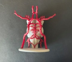
Another view of the diagram - the Circle of Willis is a collection of arteries that supply blood to the brain.

Side profile of a 3D printed diagram of the circle of Willis and the brainstem. The brainstem and the baseplate is printed in white, and from this angle the brainstem is angled backward (into the baseplate), and the muscularity and anatomical details of the brainstem is visible. The circle of Willis is printed in red, and is connected on the back of the brainstem and glued at the baseplate - it looks liek a series of small tubes circling around and up the brainstem. The model is on a black background.

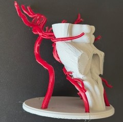
Side profile of the model!